

Ultrasound Medical Imaging
Ultrasound technology utilizes soundwaves beyond the range of human hearing to produce high-quality images without radiation exposure. Over the years, advancements in ultrasound technology have significantly improved image clarity and diagnostic accuracy.
Ultrasound is particularly effective in assessing soft tissue structures. It is a valuable tool for evaluating abdominal and pelvic organs, the breast, muscles, tendons, ligaments, thyroid gland, lymph nodes, and other small body parts.
Since ultrasound imaging is performed in real-time, it is ideal for guiding procedures such as pain-relieving injections, tendon injury treatments, and tissue sampling techniques like Fine Needle Aspiration (FNA) or biopsy.
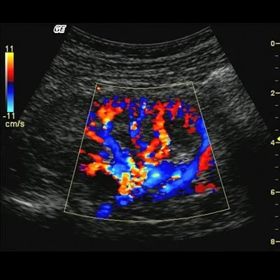
Doppler Ultrasound (Vascular Ultrasound Imaging)
Doppler ultrasound is used to visualize blood flow in arteries and veins, particularly in the limbs and neck. This non-invasive, painless imaging technique can detect vascular conditions such as narrowed arteries, varicose veins, and blood clots.
For example, Doppler ultrasound can help identify Deep Vein Thrombosis (DVT)—a condition where a blood clot reduces circulation in the veins. It is also effective in detecting arterial stenosis, or narrowing of the arteries due to atherosclerosis (“hardening of the arteries”), which commonly affects the carotid arteries in the neck and the arteries of the lower limbs.
Ultrasound Limitations
Because ultrasound waves cannot penetrate bone or gas-filled structures, it is not suitable for examining joints, bones, lungs, or the bowel. In cases where these structures require evaluation, CT scans or MRI may be more appropriate diagnostic options.
Patient Information
What is Ultrasound Imaging?
Ultrasound imaging, also known as sonography, is a safe, non-invasive medical imaging technique that uses high-frequency sound waves to produce real-time images of internal organs, tissues, and blood vessels.
Benefits of Ultrasound Imaging
✅ Non-Invasive & Radiation-Free: Unlike X-rays or CT scans, ultrasound does not use ionizing radiation, making it a safe imaging method.
✅ Real-Time Visualization: Provides live images, allowing healthcare professionals to assess organ movement and blood flow.
✅ Safe for All Patients: Ultrasound has no known risks and is suitable for all ages, including pregnant women.
Ultrasound Procedure
Before the Procedure
- In most cases, no special preparation is needed.
- Depending on the area being examined, you may be advised to follow specific instructions (see the preparation guide below).
During the Procedure
- A small handheld device called a transducer is placed on the skin over the area of interest.
- A water-based gel is applied to improve sound wave transmission.
- The transducer sends sound waves into the body, creating images displayed on a monitor.
After the Procedure
- Once the imaging is complete, you can resume normal activities immediately.
- The results will be analyzed, and your healthcare provider will discuss the findings with you.
Safety Precautions
- Radiation-Free: Ultrasound does not use ionizing radiation, making it safe for repeated use.
- Inform Your Provider: Notify your healthcare provider about any medical conditions, allergies, or recent surgeries before the procedure.
Examination Preparation Guide
Abdomen
✅ Fasting Required: Avoid eating for 6–8 hours before the scan (usually skip breakfast).
✅ Small Sips of Water Only: You may drink small amounts of water for medication intake.
Pelvis, Renal & 1st Trimester Pregnancy
✅ Full Bladder Required: Drink 1 liter of water at least one hour before the exam.
🚫 Do NOT empty your bladder until after the scan.
MSK (Limb, Joints, Muscles), Breast, Penile, Scrotum, Thyroid, Eye, Skin
✅ No Special Preparation Needed.
Vascular Doppler (Veins, Carotid, Limbs & Other Arteries)
✅ No Special Preparation Needed.
Doppler for Aorta, Renal & Spleno-Portal Axis
✅ Fasting Required: Avoid eating for 8 hours before the scan.
✅ Small Sips of Water Allowed for medication intake.
📌 Important: Please bring any previous scans (X-rays, ultrasounds, MRI, or CT) for reference.